|
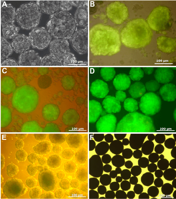
| Figure 1: Co-culture of iPS-Acta 2 derived cardiomyocytes on CultipshereS micropsheres. Empty microsphere in culture without cells (A). Coculture of eGFP+ cardiomyocytes with microspheres at day 2 post seeding (B). Coculture of eGFP+ cardiomyocytes with microspheres at day 4 post seeding (C). Manually enriched cell-loaded microsphere expressing eGFP+ at day 5-6 post seeding (D,E). MTT staining of loaded microspheres at day 8 post seeding (F). |