 |
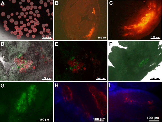
| Figure 3: Histological observation of transplanted microspheres: Cultisphere S microspheres were labeled with CMTPX dye prior to cell transplantation; Images were acquired on a fluorescence microscope (A). Viable ventricular slices prepared on a vibratome indicate the presence of CMTPX labeled microsphere observed 24h after transplantation into the left ventricular myocardium (B,C). Injected spheres were loaded with cardiomyocytes identified 24h post transplantation showing eGFP fluorescence (iPS-CMs) and red fluorescence (CMTPX labeled microspheres) (D,E). Cryo-sections of hearts transplanted with loaded microspheres showing eGFP+ iPS-CMs 24h post transplantation (F,G). One week after transplantation microsphere was visible while eGFP+ iPS-CMs were undetectable (H). After 4 weeks microspheres were degraded and no eGFP+ signals observed (I). |