 |
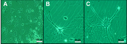
| Figure 3: Phase-contrast microscopy of SC and DRG slices cultured in NVR-Gel 8 days after vitrification. (A) Neuronal and glia migration (B) A round DRG neuron which exhibits regenerated nerve fibers, euchromatic nucleus with a prominent nucleolus. (C) A multipolar SC nerve cell with regenerated nerve fibers. |