 |
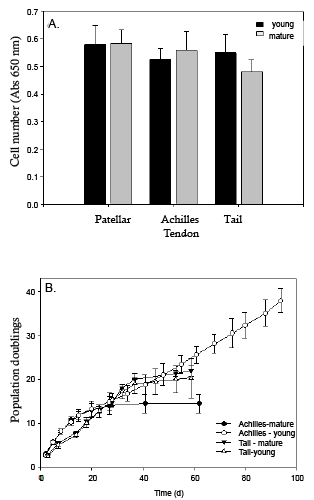
| Figure 4: Effect of age on tendon stem/progenitor cell proliferation. (A) Tendon derived cells were plated out at 1 x 10⁴ cells/cm2 and allowed to grow for 4 days, after which the cells were stopped and cell number determined by methylene blue staining. (B) Growth curves were constructed by plating tendon derived cells into T25 flasks at a concentration of 5×10³ cells/cm2 and allowing them to grow until they were 90% confluent. They were then serially sub-cultured until the cells ceased to divide. Data are presented as mean ± SEM. |