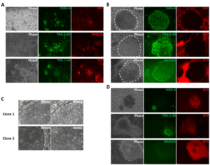
(A) Expression of SSEA-4 and RV-RFP (upper panel), TRA-1-60 and NANOG (middle panel) and TRA-1-60 and RV-RFP (lower panel) in the cell clusters / colonies on day 9 of reprogramming showing the initiation of pluripotency marker expression before the cells achieve hESC-like morphology and transgene silencing. (B) Expression of the pluripotency markers (SSEA-4, TRA-1-60 and NANOG) and RV-RFP silencing in the colonies on day 16 of reprogramming. The emerging hiPSC colonies showed characteristic hESC-like morphology and retroviral transgene silencing allowing their easy identification. (C) Higher magnification images of RV-RFPhiPSC colonies showing their hESC-like morphology- flat appearance, defined boundary and high nuclear to cytoplasmic ratio. (D) Non-hESC like RFP+ colonies which lacked the expression of pluripotency markers on day 16. White and black dotted lines indicate the boundary of hiPSC colony. All images are at 1000X magnification unless otherwise indicated. The broken lines show the characteristic boundaries of the emerging hiPSC colonies on the feeder cells.