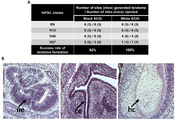
(A) Table showing the outcome of teratoma assays performed in black SCID and white SCID mice. R8, R13 and R48 are RV-hiPSC lines and N27 is an Epi-hiPSC line. (B) Haematoxylin and eosin staining of formalin fixed teratoma sections showing tissues of all the three germ layers. Representative images are shown. ne -neuroepithelium (ectoderm), he -hyaline cartilage (mesoderm) and ce-columnar epithelium (endoerm).