 |
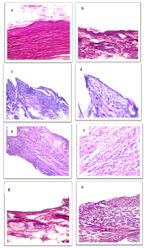
| Figure 6: H &E staining of sections from skin of female mice (a),(b) treated with BM-MSCs intraperitoneally in group 2 ,that showing full thickness healed wound and keratinized stratified squamous epithelium overlaying collagen fibers and fibroblasts.(magnification x 400).(c),(d) treated with BM-MSCs intraregionally , showing healed wound with full thickness proliferating epithelium with signs of regeneration.(e)(f) not treated with BM-MSCs , showing infected granulation tissue formed of vascular spaces , fibroblasts and mononuclear inflammatory cells.(g) not treated with BM-MSCs after one week , showing no healed wound and ulcer in the skin.(h) treated with BMMSCs after one week , showing healing wound showing basal cell proliferation covering the raw area which is early signs of healing. |