 |
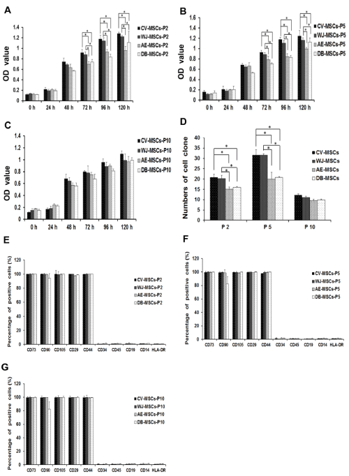
| Figure 2: Growth kinetics and characterization of four types of PDSCs. A-C: Cell proliferation of PDSCs at passages 2, 5 and 10 analyzed by WST-1 cell viability assay. Triplicate wells containing 5 × 10³ CV-MSCs, WJ-MSCs, AE-MSCs and DB-MSCs were evaluated for viability. D: Colony formation assay. Cells were seeded into 6 well-plates and the colony size and number were observed. Colony numbers that contained more than 50 cells were counted and analyzed. The data are expressed as the means ± SD of three independent experiments. * P < 0.05 compared with the control. E-G: Immunophenotypes of CV-MSCs, WJ-MSCs, AE-MSCs and DB-MSCs at passages 2 (E), 5 (F), and 10 (G) were analyzed by flow cytometry for specific cell surface markers (CD90, CD73 and CD105), hematopoietic cell markers (CD45, CD34, CD14 and CD19) and extracellular matrix receptors (CD29 and CD44) and major histocompatibility elements (HLA-DR). The data are expressed as the means ± SD of three independent experiments. |