|
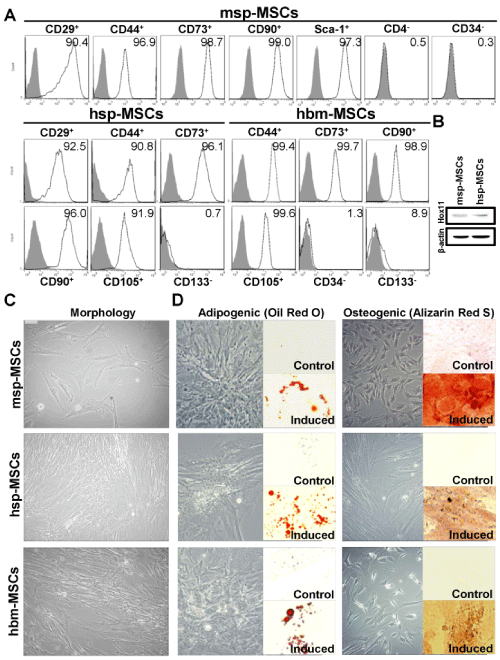
| Figure 1: Characterization of msp-MSCs, hsp-MSCs, and hbm-MSCs. (A) Expression of several stem cell markers by using flow cytometry. Representative data from three independent experiments are shown. (B) The western blot analysis was used to determine the expression of Hox11 in MSCs isolated from mouse and human spleen. (C) Representative images revealed general morphology of msp-MSCs, hsp-MSCs, and hbm-MSCs. Original magnification, 100×. (D) The multilineage differentiation capacities of hbm-MSCs, hsp-MSCs, and msp-MSCs with or without induction medium. Oil droplet stained by Oil-Red-O displayed adipogenic differentiation (left panels) at day 21. Osteogenic differentiation was evidenced by Alizarin Red staining of mineralization (right panels) at day 21. Original magnification, 400×. msp- MSCs, mouse spleen-derived mesenchymal stem cells; hsp-MSCs, human spleen-derived mesenchymal stem cells; hbm-MSCs, human bone marrowderived mesenchymal stem cells. |