|
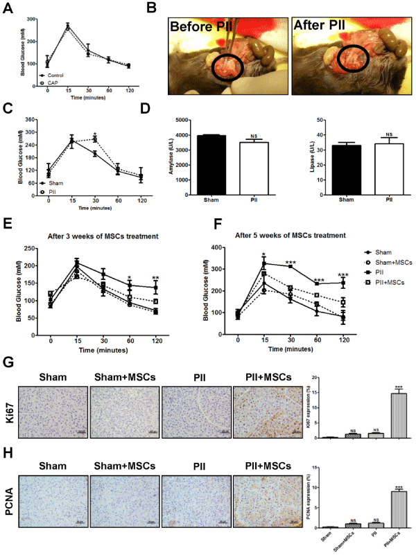
| Figure 5: Effects of msp-MSCs on glucose metabolism and cell proliferation in PII-induced mice. (A) IPGTT in mice with or without CAP. (B) Representative images showing PII mouse model induced by a vessel champ to obstruct blood flow in a part of pancreas. (C) Blood glucose values obtained from PIIinduced mice in response to IPGTT. (D) Serum amylase and lipase level were determined by activity assay. IPGTT performed in sham-operated mice and PII-induced mice transplanted with or without 1×10⁶ msp-MSCs. Blood glucose levels were measured from the snipped tail at 3 (E) or 5 weeks (F) indicated in the figures with a portable glucometer. Representative immunostaining for Ki67 (G) and proliferative cell nuclear antigen (PCNA; H) in pancreatic tissue sections. Ki67 and PCNA expression were analyzed semiquantitatively. Original magnification, 100×; scale bar, 100um. Values represent the means ± SE of three independent experiments. NS, not significant; *, P<0.05; **, P<0.01; ***, P<0.001 compared with Sham group. PII, pancreatic ischemia injury; IPGTT, intra-peritoneal glucose tolerance test; msp-MSCs, mouse spleen-derived mesenchymal stem cells. |