|
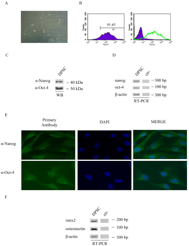
| Figure 1: a) bright-field image showing the morphology of DPSCs isolated from permanent human teeth dental pulp; b) flow cytometry analysis of cell-surface markers in DPSCs. Cells were labelled with CD146 or immunoglobulin isotype antibodies: the graph on the left shows the percentage of positive cells for CD146, on the right is evidenced the expression of CD146 (green line) respect to control; c) Nanog and Oct-4 expression in DPSCs was determined in whole-cell extract by immunoblot analysis using specific antibodies; d) expression of transcript for nanog and oct-4 genes by RT-PCR. Internal control: β-actin. Negative control: water; e) immunofluorescence staining for Nanog (green) and Oct-4 (green) in DPSCs. Nuclei were labelled with DAPI (blue). Magnification:×40; f) expression of transcript for runx2 and osteonectin genes by RT-PCR. Internal control: β-actin. Negative control: water. DPSC: dental pulp stem cells; WB: western blot; ctr-: negative control. |