 |
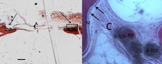
| Figure 5: a) no trabecular bone was present in the central portion of the bone defects (C). New bone extended to the basal third from the margin of the bone defect and partially surrounded the S particles (Arrows). Acid fuchsin and toluidine blue 2X, Scale bar=1mm; b) previous image at higher magnification. Traces of new bone (NB) from the periosteum were seen overlying the superficial portion of the bone defect. Osteoblasts,(black arrows) osteocytes but also osteoid (M), and blood vessels were observed. Fibrous tissue (F), inflammatory cells (I), Biomaterial (ß-TCP). Acid fuchsin and toluidine blue 100X. |