 |
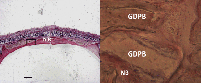
| Figure 8: a) new bone (NB) was observed surrounding some particles and extended also in central part of the bone defects. Bone formation was observed at both margins of the defect, and this was more evident at one of the margins. Acid fuchsin and toluidine blue 2X, Scale bar=1mm; b) previous image at higher magnification. New bone (NB) surrounded the biomaterial particles (GDPB). The particles were distributed along the defect and were surrounded by new bone (NB). Acid fuchsin and toluidine blue 100X. |