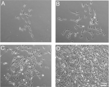
 MSCs were isolated from bone marrow at high purity. Cells expressing CD90.
1 and CD29 were identified using fluorescence-activated cell sorting. Cells
consisted of a heterogeneous cell population with a predominant spindle
morphology. Cells cultured for three days (A) and one (B), two (C), and three
weeks (D) were photographed, and MSCs proliferated to form a small colony
on day 3. A large colony of densely distributed spindle- and triangle-shaped
MSCs formed after three weeks.
MSCs were isolated from bone marrow at high purity. Cells expressing CD90.
1 and CD29 were identified using fluorescence-activated cell sorting. Cells
consisted of a heterogeneous cell population with a predominant spindle
morphology. Cells cultured for three days (A) and one (B), two (C), and three
weeks (D) were photographed, and MSCs proliferated to form a small colony
on day 3. A large colony of densely distributed spindle- and triangle-shaped
MSCs formed after three weeks.