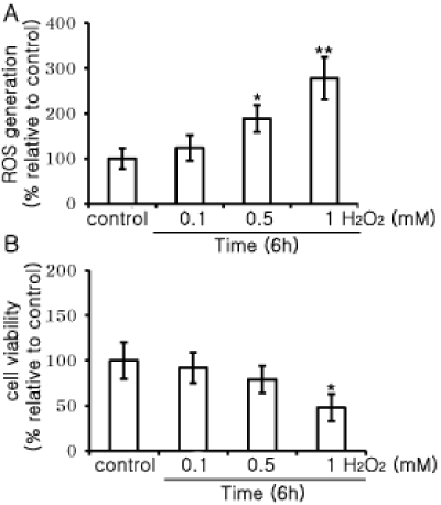
| Cells were treated with different concentrations (0, 0.1, 0.5 and 1 mM) of
H2O2 for 6 h. Cell viability was measured with an MTT assay. Cell viability
significantly decreased with increased treatment with 1 mM H2O2 at 6 h (B),
and results are expressed as the mean ± SD of fold- and percent-increases
over that of the control (n=8). ROS were measured using DCFH-DA (A).
The fluorescent intensity was measured at 530 nm after excitation at 490
nm. Fluorescent levels are expressed as percent increase over the control,
and are expressed as mean ± SD (n=8). Statistical analysis; *p<0.01, and
**p<0.05 vs. negative control (control media alone). |