|
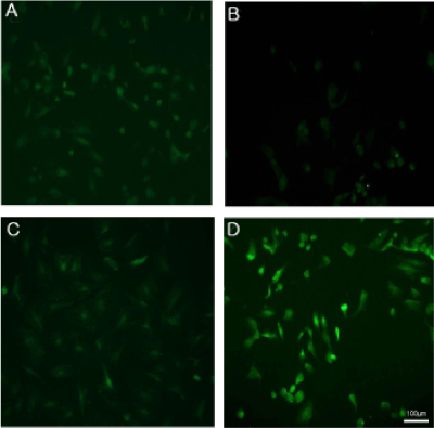
| The cellular localization of Nrf2 was determined using immunofluorescence staining with an antibody specific for Nrf2, which was fluorescently labeled using secondary antibodies. Cells were treated with 1 mM H2O2 for 6 h. Control (A), H2O2 for 6 h (B), SAM (C), and co-treatment with SAM and H2O2 (D). Representative microscopy images are shown (green). |
| Figure 8: Immunocytochemistry analysis indicates the effect of SAM on H2O2- induced Nrf2 activation in MSCs. |