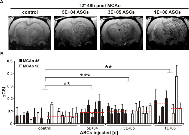
(A). Differences in ΔCSI obtained 48h post MCAo are shown for all treatment groups. Groups are controls (n = 8 for MCAo = 45 min; n = 9 for MCAo = 90 min), and animals treated with 5E+04 ASCs (n = 7; all MCAo = 45 min), 3E+05 ASCs (both n = 6 for MCAo 45 min and 90 min) and 1E+06 ASCs (n
= 4 for MCAo = 45 min; n = 3 for MCAo = 90 min). Median of each treatment group was indicated as red dotted line. Each column represents one animal with mean ΔCSI obtained in 7 brain slices per animal +/- standard deviation.
**p < 0.01 for control group vs. 1E+06 and 5E+04 ASC group, ***p < 0.001 for control group vs. 3E+05 ASC group
(B). ASC = adipose tissue derived stem cell; ΔCSI = difference in cell signal intensity; MCAo = middle cerebral artery occlusion.