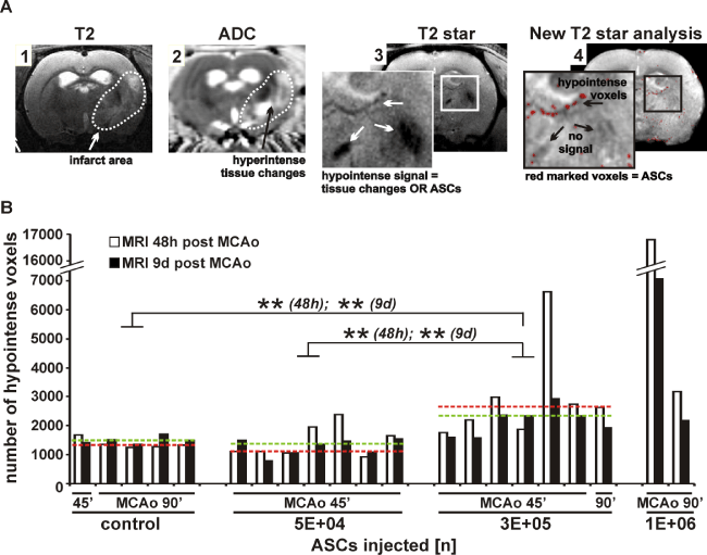
| Signal pattern changes within 9d post MCAo in infarct area (dotted line,
image 1) and consequential hyperintense signals displaying tissue changes
as shown by ADC map (image 2) are presented. Resulting ambiguous T2*-
weighted hypointense signals (image 3) were reanalysed leaving specific
ASC derived hypointense voxels (image 4) (A). Differences in voxel number
obtained 48h and 9d post MCAo are shown for all treatment groups. Groups
were controls (n = 1 for MCAo = 45 min; n = 4 for MCAo = 90 min), and
animals treated with 5E+04 ASCs (n = 7; all MCAo = 45 min), 3E+05 ASCs
(n = 6 for MCAo = 45 min; n = 1 for MCAo = 90 min) and 1E+06 ASCs (n = 2
for MCAo = 90 min). Median for each group was indicated as red dotted line
for voxel numbers obtained 48h post MCAo and as green dotted line for voxel
numbers obtained 9d post MCAo. Each pair of columns represents values
of one animal and two MRI time-points. **p < 0.01 for 3E+05 ASC group vs.
control group and 5E+04 ASC group (B). ASC = adipose tissue derived stem
cell; MCAo = middle cerebral artery occlusion. |