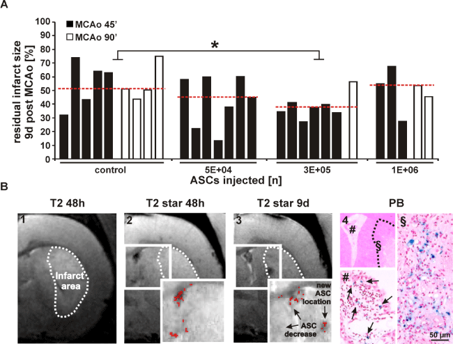
Differences in residual infarct volumes obtained 9d post MCAo are shown for all treatment groups. Groups are controls (n = 5 for MCAo = 45 min; n = 4 for MCAo =
90 min), and animals treated with 5E+04 ASCs (n = 7; all MCAo = 45 min), 3E+05 ASCs (n = 6 for MCAo 45 min; n = 1 for MCAo = 90 min) and 1E+06 ASCs (n = 3
for MCAo = 45 min; n = 2 for MCAo = 90 min). Median for each group was indicated as red dotted line. Each column represents residual infarct size for one animal
assessed as percentage of individual infarct size obtained 48h post MCAo for each individual animal. *p < 0.05 for 3E+05 ASC group vs. control
(A). Migration activity in the ischemic hemisphere (image 1, dotted line) of one animal treated with 3E+05 ASCs is shown. A strong hypointense signal in the right
ventricle of the appending T2*-weighted image (image 2) obtained after 48h decreases within 9d post MCAo with a new hypointense spot appearing in striatum close
to infarct area (image 3). Signals are confirmed by voxel analysis. Histological analysis of the same location reveals positive PB staining (image 4, location # and §)
(B). ASC = adipose tissue derived stem cell; MCAo = middle cerebral artery occlusion; PB = Prussian blue. |