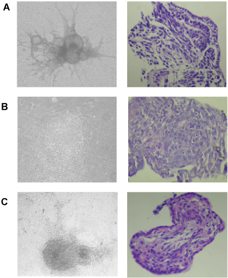
| A, B, and C are from EBs at 14 days after in vitro differentiation showing
typical morphology and histology characteristics of differentiated tissues
from the three germinal layers. (A) Neural epithelium characteristics in an
ectoderm layer. (B) Mesenchymal characteristics in a mesoderm layer. (C)
Pseudostratified columnar epithelium characteristics in an endoderm layer.
Phase contrast images (left panels) are shown with 10X magnification.
Haematoxylin and Eosin stained images (right panels) are shown at 60X
magnification. |