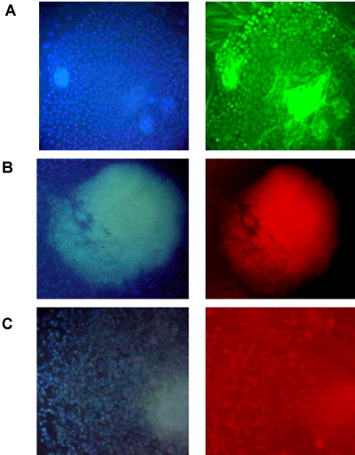
| A, B, and C are from EBs at 14 days after in vitro differentiation. (A) EB
showing βIII-tubulin positive expression (green) which is characteristic of
neuroectoderm differentiation. (B) EB showing myosin positive expression
(red) which is characteristic of cardiac muscle (mesoderm differentiation).
(C) EB showing GATA positive expression (orange) which is characteristic of
endoderm differentiation. Nuclei were visualized with DAPI stain (blue) (left
panels). Fluorescence images are show in magnification of 60X. |