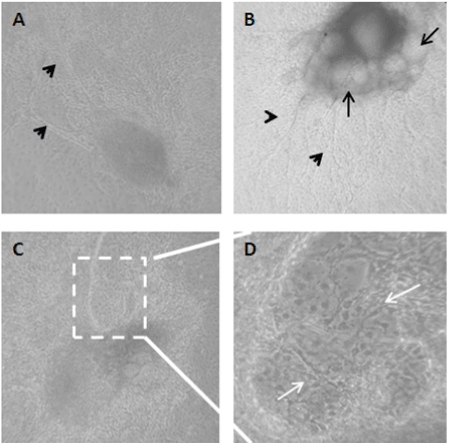
| (A-B) Neural progenitor spheres with extensive cell growth around the clusters
and neurite grew radially from the middle EB sphere (black arrow heads). (B)
Neural rosettes are observed inside the floating spheres (black arrows). (C)
Neural rosette with high confluence of early progenitors that appear after 3
weeks of in vitro differentiation from the H1 ES cell line. (D) Boxed region from
C panel, shown in 60X magnification and displays neuronal generation in the
outgrowth area. Cells generated are in migration status (white arrows). Phase
contrast images (A-C) are at a magnification of 10X. |

