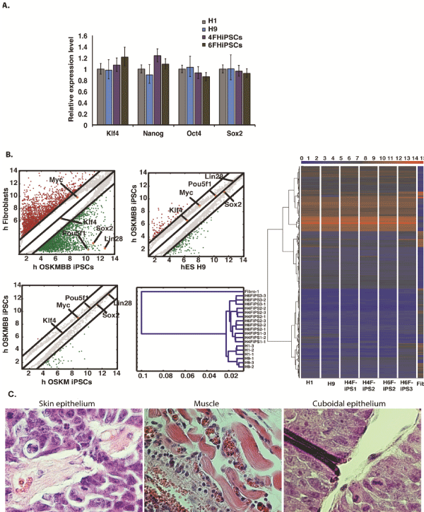
| (A) Quantitative PCR analysis for pluripotent markers in H1 human ESCs, H9 human ESCs, OSKM human iPSCs and OSKMBB hiPSCs. The expression levels are relative to H1 human ESCs using primers specific for endogenous transcripts, and on the logarithmic scale. Transcript levels were normalized to Gapdh expression. (B) Correlation plot analysis of human fibroblasts and OSKMBB hiPSC (upper left), OSKMBB hiPSCs and H9 (upper right) OSKMBB hiPSCs and OSKM hiPSCs (lower
left). The black lines indicate twofold differences in gene expression levels between the paired cell populations. The transcript expression levels are on the log2 scale. Hierarchical cluster analysis (lower right) heat map (right panel) analysis of global gene expression from human fibroblasts, H9 human ESCs, H1 human ESCs, OSKM hiPSCs (H4FIPS) and OSKMBB hiPSCs (H6FIPS). The abscissa numbers in the hierarchical cluster correspond with the standardized Euclidean distance. A color bar (top) indicates the color code gene expression in log2 scale. (C) Teratoma formation after 6–8 weeks transplantation of OSKMBB human iPS cells into SCID mice. Teratomas were sectioned and stained with haematoxylin and eosin at 6–8 weeks. Histological section of identified cells representing all three germ layers: ectoderm (skin epithelium), mesoderm (skeletal muscle) endoderm (gut epithelium). |