|
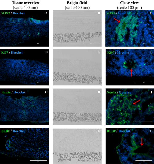
| Primary antibodies (green) were SOX2 (A, C), Ki67 (D, F), Nestin (G, I), Nuclei were stained with Hoechst (blue). The first column is a “Tissue overview” (scale 400 μm), the second column is a “Bright field” showing the position of the tissue on the scaffold. The third column shows a close up view of the tissue, where a red arrow marks neural tube-like structures (NTLS). |
| Figure 1: Immunohistochemistry (IHC) staining of human induced pluripotent stem cells differentiated in a scaffold into 3D neural tissue. |