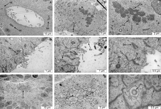
| Images of ultrastructural components resembling the developing neural tube. A (scale bar 10 μm): overview of the NTLS lumen (L) with periluminar mitoses (Mi). B
(scale bar 2 μm): Close view of mitosis (Mi) next to lumen (L) with visible tight junctions (Tj) at apical cell lining. C (scale bar 2 μm): Periluminar mitosis (Mi) with visible
centrioles (Ce). D (scale bar 1 μm): Luminal epithelial lining with basal bodies (Bb) and tight junctions (Tj). E (scale bar 1 μm): Luminal epithelial lining with cilia (Ci)
and microvilli (Mv). F (scale bar 0.5 μm): Luminal epithelial lining with basal bodies (Bb) and tight junctions (Tj). G (scale bar 10 μm): Luminal tangential cut (Lt) with
surrounding nuclei (N). H (scale bar 2 μm): Closer view of luminal tangential cut showing presence of primary cilia (Pc) and basal bodies (Bb). I (scale bar 0.5 μm):
Close up view of luminal tangential cut showing primary cilia (Pc) and tight junctions (Tj). |