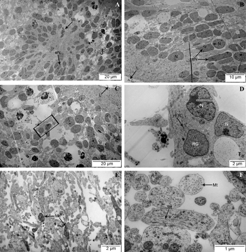
| A (scale bar 20 μm): NTLS overview with the notion of close by lumen showing mitosis (Mi) and elongated nuclei (eN). B (scale bar 10 μm): Periluminar NTLS view
with visible lumen (L), elongated nuclei (eN) and round nuclei (N). C (scale bar 20 μm): Periluminar NTLS view with visible lumen (L) and radial glia-like cell (black box)
with elongated cytoplasm radiating towards lumen. D (scale bar 2 μm): Close up view of radial glia-like cell (RG) with visible rough endoplasmic reticulum (rER), seen
in figure C, with another cell positioned close to its nucleus (N). E (scale bar 2 μm): Mesh of axon-like cellular extensions seen in the surrounding tissue. Here cut at
varying orientation with mitochondria (Mc) present. F (scale bar 1 μm): Close view of axon-like cellular extensions with microtubules (Mt) seen in different orientations. |