|
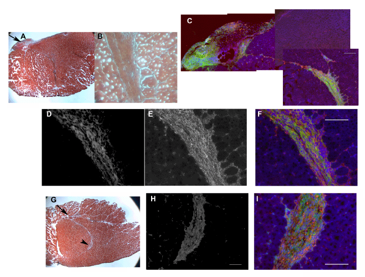
| Figure 7: pCPCs transplanted in the heart did not express cardiac troponin and were found in fibrotic tissue. (A) Transplanted pCPCs identified as GFP+/ PKH26+ cells (green and red fluorescent, respectively) were located in the left ventricular wall. The bright red florescence emitted from PKH26 greatly facilitated the screening of the heart tissue to locate the transplanted cells. (B) H&E staining showed fibrotic-like tissue in the transplanted area. (C) Higher magnification of area pointed by an arrow in (B). (D) Massons trichrome staining showed collagen deposition (in blue) in the transplanted area. (E) Higher magnification of area pointed by an arrowhead in (D). (F) Higher magnification of area pointed by arrow in (D). (G-J) Transplanted pCPCs identified as GFP+/PKH26+ did not exhibit cardiomyocyte morphology and did not stain positive for cardiac Troponin T. (K-N) Another transplanted heart clearly showed GFP+/PKH26+ cells in the injection track were negative for cardiac Troponin T (M-N insets). Scale bars = 100 µm. |