|
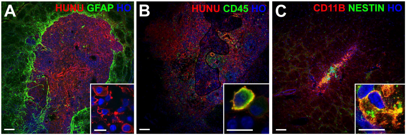
| Figure 3: Rat brain sections with dead graft cell debris. Panel A shows dead graft cell debris with the red-labeled human nuclear marker (HUNU) surrounded by reactive astrocytes labeled green for GFAP. The inset of panel A shows that the HUNU marker, which should only be in the nuclei of intact graft cells, was instead found in the cytoplasm of cells invading the graft area with a morphology consistent with activated microglia. Panels B and C show grafts with areas of live graft cells with intact nuclei as well as areas of dead graft cell debris. Panel B shows cells with the morphology of activated microglia having cytoplasm that co-labeled for HUNU in red and the leukocyte marker CD45 in green. Panel C shows cells with cytoplasm that co-labeled for the microglial marker CD11B in red and a human-specific antibody for the neural progenitor marker nestin in green. All nuclei are labeled blue with Hoechst (HO); scale bars = 100 um (10 um for the insets). |