|
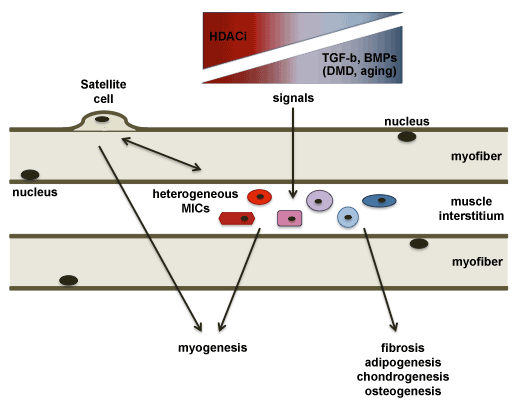
| Figure 1: Different cell types located in the interstitium of skeletal muscles are depicted and indicated as muscle interstitial cells (MICs). These cell type share a partial pluripotency within the mesoderm-derived lineages and can contribute to the repair of skeletal muscles directly or indirectly. Indirect interactions with other cell types, and in particular with the main cellular effectors of muscle regeneration (the satellite cells), has been shown to promote early regeneration events in injured muscles, possibly via paracrine signals. Given their potential to adopt a fibro-adipogenic or myogenic phenotype in response to environmental cues, MICs can be a pivotal cellular component that determines whether muscle healing occurs by regeneration or fibro-adipogenic degeneration. As such, MICs could contribute to the pathogenesis of chronic degenerative muscular diseases, as well as to age-dependent decline of muscle mass and activity (sarcopenia). Signaling pathways, such as transforming growth factor beta (TGF-b) and bone morphogenetic protein (BMP) signallings, that promote degenerative outcome of failed skeletal muscle regeneration during aging and Duchenne Muscular Dystrophy, are depicted. Given the phenotypic and functional heterogeneity of MICs, the identification of sub-populations and their dynamic transitions in response to developmental, physiological and pathological signals, based on transcriptome analysis at the single cell level, will clarify their function and identify potential target for their pharmacological manipulation, for example by HDACi treatment that can reverse skeletal muscle degeneration [33]. |