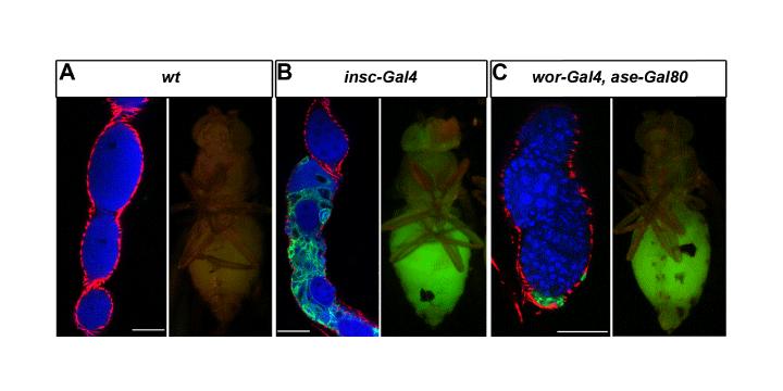
(A) insc-Gal4 driver control,
(B) insc-Gal4 driven numb RNAi and
(C) wor-Gal4, ase-Gal80 driven numb RNAi. In (B and C) the presence of GFP-labeled cells (in green) in the ovarioles indicates the presence of metastasis. DNA is labeled with Toto-3 iodide (in blue) and muscle layers by phalloidin (in red). Scale bars are 30 µm.