|
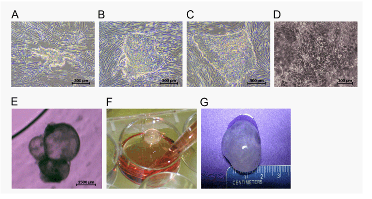
| Figure 1: Corneal orb differentiation from pluripotent human stem cells. A. Early appearance of pluripotent stem cell colonies that will go on to form corneal orbs. The small, tightly packed cells in the colony are distinguishable morphologically from the spindle-shaped feeder cells. Scale bar = 300 µm. B. Proliferation of the pre-cornea colonies results in larger colonies of irregular shapes but with clear edges demarcating the colony from spindle-shaped feeder layer cells. Scale bar = 300 µm C. Another expanding colony of differentiating cells easily distinguishable by light microscopy from the feeder layer. Scale bar = 300 µm D. Higher power appearance of differentiated pluripotent cells just before the cells are passaged onto low-adherence, deep plates. Scale bar = 100 µm E. Differentiating hpSC from which small (˜1-2 mm) orbs have emerged. Scale bar = 1500 µm F. Mature corneal orb after 120 days of differentiation is translucent and fluidfilled. G. Large, mature corneal orb after 120 days of differentiation with diameter of ˜15mm. |