|
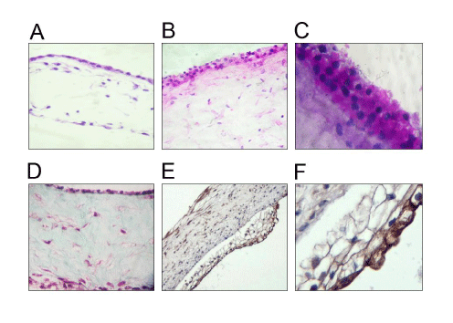
| Figure 2: Characterization of multilayered human pluripotent stem cell-derived corneal orbs. A. H&E stain of differentiating corneal orb at ~50 days of differentiation (10X) shows the largest component of corneal construct, the stromal layer, below a single layer of epithelial cells (top of figure). B. H&E stain of sectioned corneal orb at the end of 120 days differentiation, showing expansion of the epithelial layer (10X) C. PAS staining of corneal orb at the end of differentiation shows accumulation of glycogen or other polysaccharides in the epithelium during differentiation (20X). D. Trichrome staining of corneal orb at end-differentiation shows accumulation of stromal collagen (blue), the major protein of normal corneal stroma. Fibrin strands (pink) are also seen surrounding nuclei of presumed differentiating keratocytes (10X). E. After exposure to an air-liquid interface, the epithelial layer expands. Brown staining is for cytokeratins (10X). F. Higher magnification of (E) emphasizes intense cytokeratin staining of the most superficial epithelial cell layer (20X). |