 |
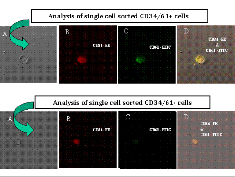
| Figure 1: Confocal microscopy of single sorted CD34+/CD61+ and CD34+/ CD61- cells. Legend: Microscopy of CD34+/CD61+ and CD34+/CD61- sorted single cells (picture A). Sorted cells were analyzed at the single cell level as illustrated by one representative experiment which show CD34PE (red) stained cells (picture B), CD61FITC differential stained cells (picture C) and combined CD34PE & CD61FITC stained cells (picture D). |