|
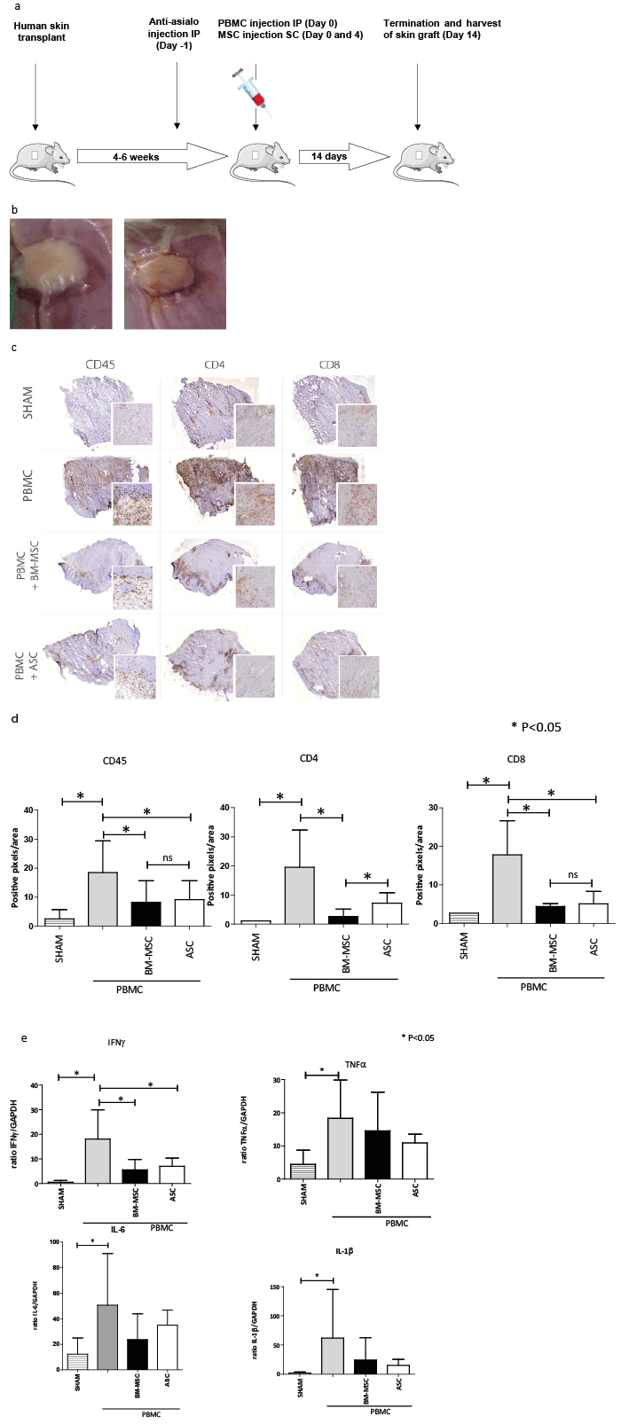
| Figure 3: (A) HuSCID mouse model: human skin is transplanted; mouse NK cells are depleted using anti-asialo GM1 injection at day-1; allogeneic PBMC are injected intraperitoneal at day 0; MSC are injected on day 0 and day 4; and after 14 days, mice are sacrificed and skin grafts are harvested. (B) Representative example of engraftment process of skin graft, pictures taken at 12 and 16 days after skin graft transplantation. (C) Immunohistological evaluation of explanted skin grafts. Staining for CD45+, CD4+ and CD8+ T cells of skin grafts explanted from mice receiving only a skin transplant (SHAM group); mice receiving skin graft and PBMC; mice receiving skin graft, PBMC and BM-MSC; and mice receiving skin graft, PBMC and ASC. Representative biopsies are shown. (D) Quantitative evaluation of CD45+, CD4+ and CD8+ cells in biopsies of study groups. Data represent mean (SD), *indicates P<0.05. (E) mRNA gene expression in explanted skin grafts of IFNγ, TNFα, IL-6 and IL-1β of study groups. Ratio gene/GAPDH x1000 is shown. Data represent mean (SD), *indicates P<0.05. |