 |
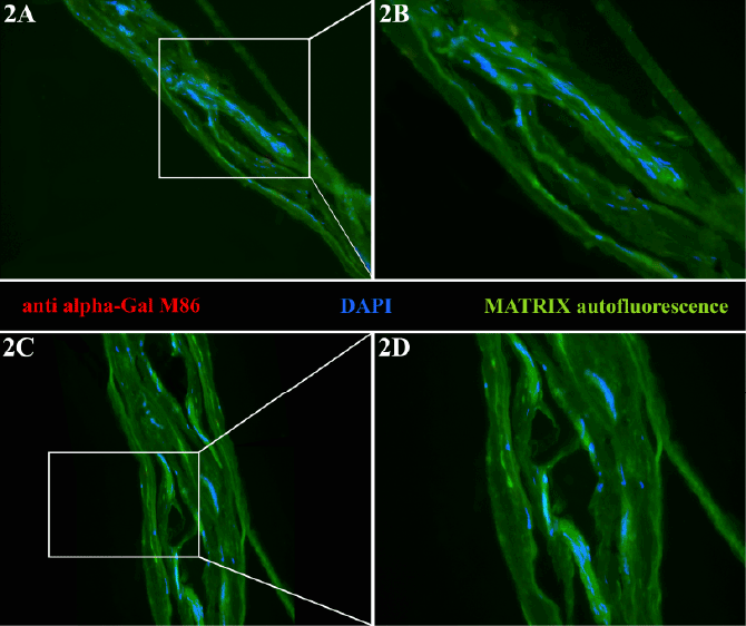
| Figure 2: Alpha-Gal epitope distribution in two different samples (A-B and C-D) of decellularized CorMatrix SIS-ECM material by immunofluorescence analysis. Note the presence of nucleic acid remnants in the declared acellular scaffold. Magnification 20× for A and C; magnification 40× for B and D. Antialpha- Gal monoclonal antibody M86 - red; Nucleic acid material - blue; Matrix autofluorescence - green. |