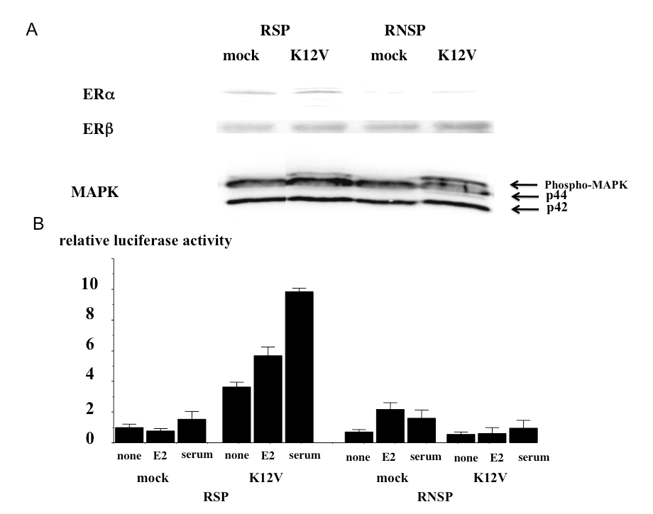
A) The expression levels of ERα and β were analyzed by Western blot. The level of ER β protein was enhanced in RSP and RSP-K12V cells compared with the levels in NSP and NSP-K12V cells. The level of ERβ was constant across all cell types. The total MAPK level is shown as an internal control. Although the levels of total MAPK were similar in all of the cell types, activation of MAPK (upper band denoted by the arrow) was observed in RSP-K12V cells and RNSP-K12V cells, but not in RSP cells or RNSP cells. Representative data are shown. Similar results were obtained in three independent experiments. The ER transcriptional activity in each of cells type by a luciferase reporter assay using luciferase reporter plasmid containing ERE-sequence in the promoter region in the presence or absence of 10-7 M E2 and/or 10% serum. The ratio of luciferase activity to RSP-mock cells in the absence of both 10-7 M E2 and 10% serum was calculated. ER activity in all conditions was significantly enhanced in RSP-K12V cells compared with that in RSP-mock cells, RNSP-mock cells and RNSP-K12V cells (p < 0.05). Data are represented as the means ±SEM from three independent experiments.