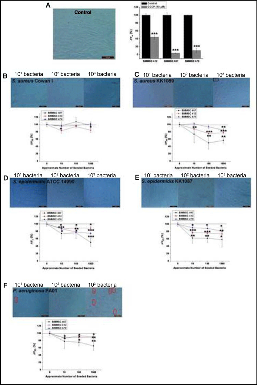
 A) Trypan blue staining of control cells and response to CCCP
(protonophore) that was used as a positive control.
A) Trypan blue staining of control cells and response to CCCP
(protonophore) that was used as a positive control.B) S. aureus Cowan I caused slight depolarization of ΔΨm.
C) S. aureus KK1089 induced changes in ΔΨm and Trypan blue after 4 hours.
D) S. epidermidis ATCC 14990 and
E) S. epidermidis KK1087 induced depolarization of ΔΨm
F) Depolarization of ΔΨm and Trypan blue staining at 4 hours in ΔΨm PA01 infected BMMSCs The fluorescent 595nm / 535 nm - ratio of JC-10 was used to determine ΔΨm and the 595 nm / 535 nm - ratio of infected BMMSC were compared to uninfected control cells. Results are indicated as mean ± SD of four independent replicates. Magnification power was 200x in all panels. Red circles indicate Trypan blue positive nuclei. Black squares indicate the S .aureus produced aggregates produced by S. aureus.