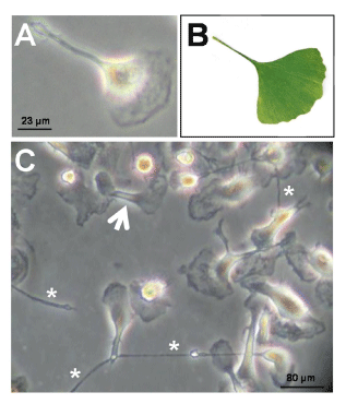
A. High-magnification view showing the characteristic cell morphology with a broad cytoplasmic extension at one pole of the cell and a thin cytoplasmic process at the opposite pole.
B. Gingko biloba leaf, to which the cell shape has been compared.
C. Cells after 50 days in culture (arrow, elongated cell process; asterisks, cytoplasmic filaments apparently establishing intercellular contacts).