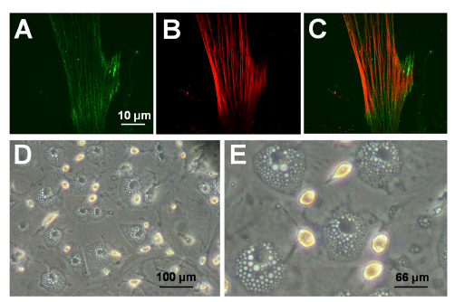
A. Immunostaining for sarcomeric a-actinin with Alexa 488 (green). Nuclei were stained with DAPI (blue).
B. Immunostaining for a-SMA with Alexa 594 (red) and DAPI (blue).
C. Merge. D/E. Phase photomicrographs of cardiac-derived cells showing adipogenic differentiation, as evidenced by intracytoplasmic fat droplets (low and high magnification views, respectively).