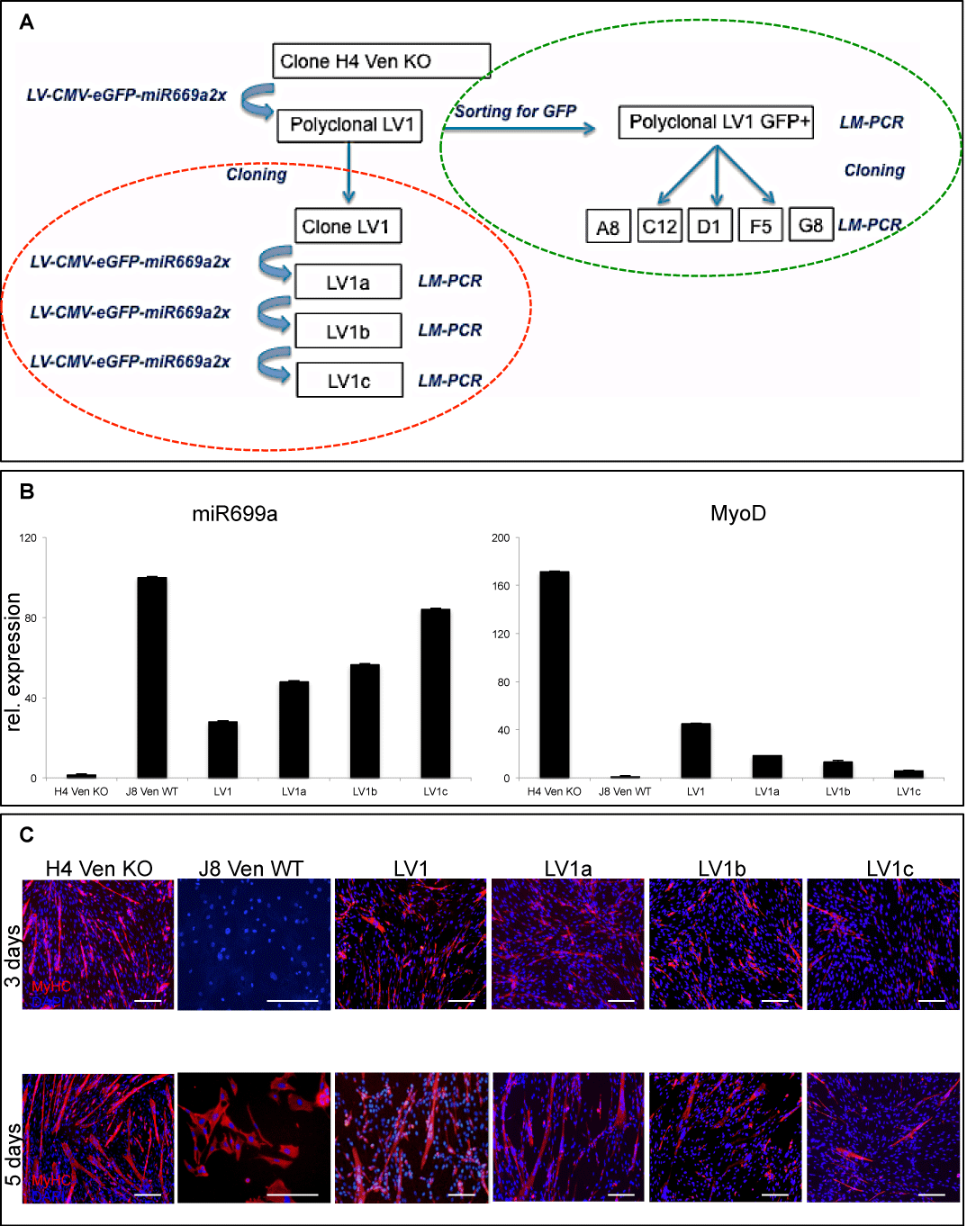
 (A) Flowchart of
experimental plan. We adopted two different approaches to evaluate the integration site profile of LV-CMV-eGFP-miR669a2x vector in infected cardiac progenitors by
LM-PCR. 1) Analysis of vector integrations in polyclonal Sgcb-null cells upon multiple infections (red dashed circle). 2) Analysis of vector integrations in monoclonal
infected clones (A8, C12, D1, F5 and G8), derived from GFP+ Sgcb-null cardiac progenitors previously transduced with the same viral vector (green dashed circle).
Note that the protocol of infection and MOI have been standardized for all experiments.
(A) Flowchart of
experimental plan. We adopted two different approaches to evaluate the integration site profile of LV-CMV-eGFP-miR669a2x vector in infected cardiac progenitors by
LM-PCR. 1) Analysis of vector integrations in polyclonal Sgcb-null cells upon multiple infections (red dashed circle). 2) Analysis of vector integrations in monoclonal
infected clones (A8, C12, D1, F5 and G8), derived from GFP+ Sgcb-null cardiac progenitors previously transduced with the same viral vector (green dashed circle).
Note that the protocol of infection and MOI have been standardized for all experiments. (B) TaqMan assay analysis for miR669a expression (left panel) and qPCR analysis for MyoD expression (right panel) in wt and Sgcb-null clone after a single infection (LV1) with LV-CMV-eGFP-miR669a2x or multiple infections (LV1a, after double infection; LV1b, after three infections and LV1c after four infections) using the same protocol. MyoD expression decreases in a dose-response manner according to miR669a increment.
(C) Immunofluorescence analysis of differentiating Sgcb-null (H4 Ven KO, LV1, LV1a, LV1b and LV1c) and wt (J8 Ven WT)cardiac clones, at 3 and 5 d after serum starvation (2% HS). Skeletal muscle differentiation was impaired by miR669a transduction in dose dependent manner. Note that only few myocytes are present in multiple infected clones compared to controls, after 3 and 5 days from serum starvation; scale bar = 100 μm.