|
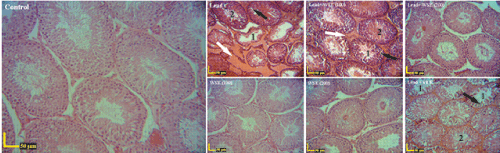
| Figure 1: Histological structure of rat testis using H&E staining. Inactive and degenerative seminiferous tubules (1), edema (white arrows), discontinuity and vacuolation changes (black arrows) were seen in seminiferous tubule epithelium in groups which received lead, compared to those in control group. Active and inactive seminiferous tubules and edema were seen in combined lead and WSE (100 mg/kg/day), and combined lead and vitamin E groups. Combined lead and WSE (200 mg/kg/day), WSE (100 mg/kg/day) and WSE (200 mg/kg/day) groups showed normal activity. Number 2 shows active seminiferous tubules. Values in parentheses are shown in mg/kg/day. |