(a)-(c), AFM topography, peak force error, and 3D topography of bovine articular cartilage in ultrapure water. Black arrows in (b) point to thin fibrils.
|
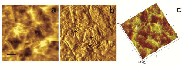
| Figure 2: Fluid AFM images showing two populations of fibril diameters
at 30 mm-deep layer of articular cartilage of normal bovine (scale bars,
500 nm). (a)-(c), AFM topography, peak force error, and 3D topography of bovine articular cartilage in ultrapure water. Black arrows in (b) point to thin fibrils. |