|
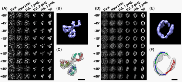
| Figure 2: 3D reconstructions of a IgG antibody and a 17nm HDL by IPET (6). (A) A single IgG antibody was imaged by OpNS tomography. By IPET, the images were aligned, and (B) reconstructed into a 3D density map at resolution of ~14 Å. (C) Each domain of 3D map can near-perfect fit to the crystal structure domain in size and shape. (D) Using IPET, a 3D map of an individual 17nm HDL particle can be reconstructed. (E) 3D reconstruction at resolution of ~36 Å indicated a ring-shape structure of apoA-I. (F) The ring length suggested three apoA-Is within 17nm HDL. Scale bars, 5 nm. |