|
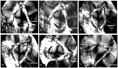
| Figure 3: A group of birefringent vortices is revealed in the collagen bundles after rotating the microscope stage while keeping the polariser-analyser PPLs crossed and motionless. Rotation of the microscope stage causes changes in the angles of collagen fiber axes relative to the PPLs and affects the fibres’ birefringence intensity. The images from panels a to f depict the spiral path of fibers around a center and indicate rotation of the polarized light caused by bundled fibers. Comparing these images sequentially, variation of the birefringence intensity is by grey levels. The fibers in the northeast region of panel a are not observed because they are oriented in an extinction position. Following stage rotation, these fibers show birefringence (d) because they become positioned at 45° relative to the polarizer-analyzer PPLs. The panels also show partial presence of other vortices for collagen bundles that emerge differently at the level of the section. The vortex images are surrounded by the endotendon limits. Bars = 100 μm. |