|
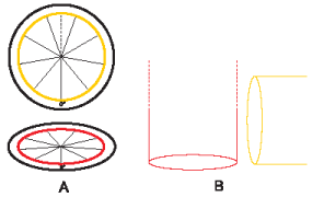
| Figure 3: Centrosome theoretical geometrical model: a spherical reference system composed of two orthogonal protractors/goniometers. A: frontal view of two orthogonal protractors/goniometers, subdivided into nine sectors, which schematizes the two orthogonal centrioles: the first (horizontal) represents the base of the MC, arranged on the equatorial “x y“ plane; its “0° ” mark is used to orient the protractor/centriole; the second, the DC (vertical, orthogonal to the first), is closer to the reader: both “0°“ marks coincide; it is convenient to consider the second protractor divided, by its “vertical” diameter crossing the “0°” mark, into two halves (two opposite symmetrical hemiprotractors). B: schematic lateral view of the proximal end of both centrioles (during S, G2) to show the respective position of the above two sections. (From: M. Regolini Centrosome: a geometrical model Lambert Academic Publishing Germany 2014) |