 |
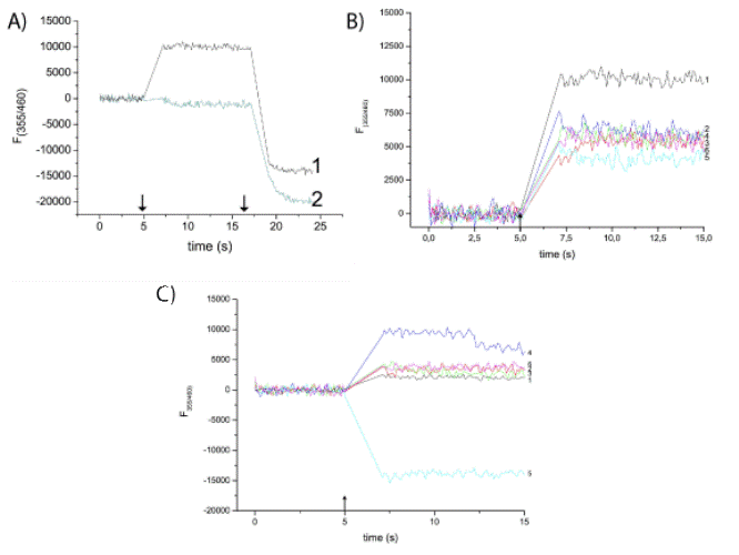
| Figure 4: Optical analysis of MjK2 activity. A) Liposomes were reconstituted with purified MjK2 (MjK2 channelosomes) (1) or control liposomes without MjK2 protein (2) contain inside 1 M KCl, 20 mM Na-citrate pH 4. B) MjK2 channelosomes contain inside 1, 0.1 or 0.01 M KCl,1 M glycine and 20 mM Na-citrate pH4 (traces 1-3, respectively) or 1 M glycine and 20 mM Tris pH8 (traces 4-6, respectively). C) MjK2 channelosomes contain inside 20 mM Na-citrate pH4, 1 M KCl and 25 mM MgCl2, pH 4 (trace 1), pH 8 (trace 2), 25 mM CaCl2 (traces 3 (pH 4), 4 (pH 8), respectively) or 0.1 mM cAMP (traces 5 (pH 4), 6 (pH 8), respectively). For optical flux measurements, 5 µl samples (A-C) were transferred into a black microplate containing an external solution of 1 M glycine, 10 mM Tris-HCl, pH8, 10 mM BaCl2 and 10 µM PBFI at pH 8. After 5 s (arrow), 10 µL of EDTA (50 mM) was injected into the running experiment using a sample rate of 0.1 s-1. After 10 s (arrow, Figure. 5A), an additional injection of 10 µL followed, and the reaction was monitored for additional 10 s. For comparison, the data were normalized to the baseline using the first 5 s with fluorescence intensities of approximately 7-9x10⁴ counts. |