|
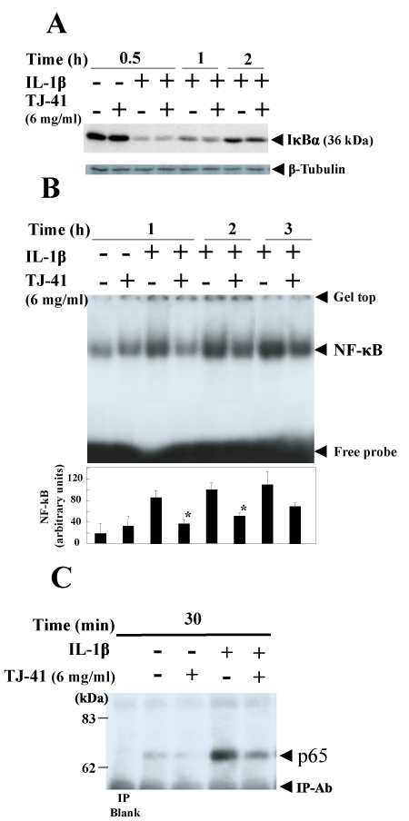
| Figure 3: Effects of TJ-41 on the degradation of IκB protein and activation of NF-κB. Cells were treated with IL-1β (1 nM) in the presence or absence of TJ-41 (6 mg/ml) for the indicated times. A: Cell lysates (20 µg of protein) were subjected to SDS-PAGE in a 12.5% gel, followed by immunoblotting with an anti-IκBα or anti-β-tubulin antibody. B: Activation of NF-κB. Nuclear extracts (4 μg of protein) were analyzed by EMSA (upper). The bands corresponding to NF-κB were quantified by densitometry (lower, means ± SD for n=3 experiments; *P<0.05 vs. IL-1β alone). C: Nuclear translocation of NF-κB subunit p65. Nuclear extracts were immunoprecipitated, and the immunoprecipitates were analyzed by western blotting with an anti-p65 antibody. |