 |
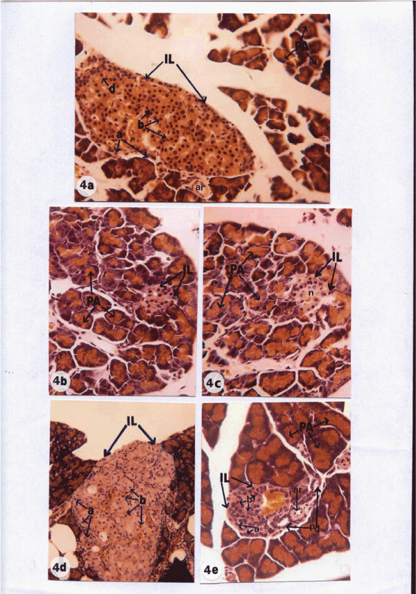
| Figure 4: Photomicrographs of sections of pancreas of normal (Figures 4a), diabetic control (Figures 4b and 3c) and diabetic treated (Figures 4d and 4e) female rats. Normal rats showed intact islets of Langerhans (IL) with alpha cells (a) at the periphery, beta cells (b) in the core and delta cells (d) adjacent to beta cells with enlarged size. Diabetic rats showed a deleterious decrease in the size of islets (Figures 4d and 4e) with necrosis (n). Diabetic rats treated with 4b exhibited marked increase in the size of islets and number of islet cells (a and b). Pancreatic ductule (bd) and pancreatic artery (ar) are also visible. |