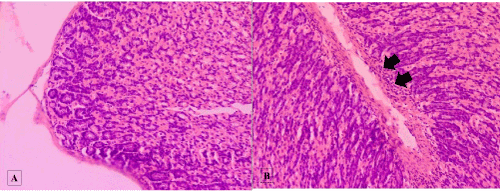
Left Panel (A): Normal stomach in non-diabetic groups.
Right Panel (B): In all of the diabetic groups, cell infiltration (arrow) were observed: Magnification 400x.
|
| Figure 3: Shows sections of the stomach - stained by H&E Left Panel (A): Normal stomach in non-diabetic groups. Right Panel (B): In all of the diabetic groups, cell infiltration (arrow) were observed: Magnification 400x. |