|
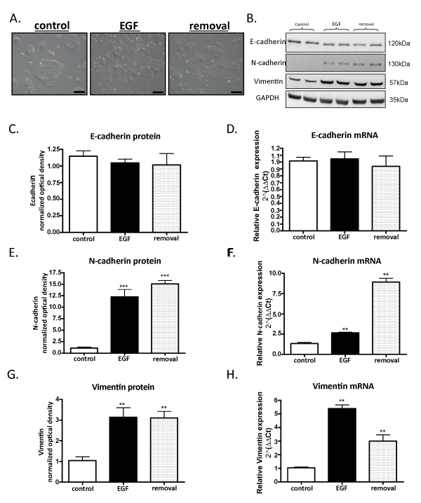
| Figure 1: Long term incubation with Epidermal Growth Factor (EGF) causes persistent change in OVCA 433 cell morphology and induction of mesenchymal markers A) Representative phase images taken with 10X objective of OVCA 433 control, long term EGF-treated (EGF) and long term EGF treated cells in which the ligand was removed for at least 7 days (removal). Scale bar=200 μm. B) Representative western blots of E-cadherin, N-cadherin and vimentinfor control, EGF and 7 day removal (removal) samples with GAPDH as a loading control C) Quantification of normalized optical densities for E-cadherin in control, EGF and removal samples D)Relative E-cadherin mRNA expression. E) Quantification of normalized optical densities for N-cadherin in control, EGF and removal samples showed a significant increase in N-cadherin protein in EGF and removal samples, n=5, ***p<0.001. F) Relative N-cadherin mRNA expression was also increased in EGF and removal samples, n=5, **p<0.01. G) Quantification of normalized optical densities for vimentin in control, EGF and removal samples showed a significant increase in vimentin protein in EGF and removal samples, n=5, **p<0.01. H) Relative vimentin mRNA expression was also increased in EGF and removal samples, n=5, **p<0.01. |